The place of 2D/3D/4D ultrasound in gynecology and obstetrics
Ultrasound examinations during pregnancy: when they are needed and what they reveal
Pregnancy becomes visible by ultrasound after 3-4 weeks after fertilization. This is usually 7-14 days from the expected menstrual cycle. In that interval, it is necessary for the future mother to come for an ultrasound examination, which will show the existence of pregnancy, the number of embryos, the baby’s heartbeat, as well as the most important information, which is where the pregnancy is developing, whether the fetus is in the uterus or outside the uterus.
The next important ultrasound examination is performed from the 12th to the 14th week of pregnancy. Then the development of the fetal morphology of the head, face, heart, abdominal and spinal wall, bladder, stomach, extremities is examined in detail, and then the sex can be determined for the first time during pregnancy.
Based on the measurement of the size of the fetus, the exact date of delivery can be determined. Special emphasis is placed on the determination of important parameters, first of all the presence of the nasal bone and the size of the cervical fold (NT), and then on sonoembryology – the morphological characteristics of the fetus and small ultrasound markers, which can indicate whether there are chromosomal or congenital anomalies.

After the ultrasound examination, the mother’s blood is sampled in the laboratory on the same day to perform a prognostic double-test. It represents a biochemical screening, that is, it shows a unified finding of measured ultrasound parameters and values of biochemical markers from the mother’s blood for the risk of giving birth to a newborn with Down’s, Patau’s or Edwards’ syndrome. This examination is recommended for all pregnant women, regardless of age. It represents a basic ultrasound examination that will direct further prognostic or diagnostic procedures.
In cases where a pregnant woman misses this testing period. Pregnant women can undergo an ultrasound with biochemical screening in the second trimester, a triple or quadruple test, but their reliability is much lower than the double test. They are useful in cases where the double test was not done on time.
An ultrasound examination of the detailed development of the fetal anatomy is best performed in the period between the 20th and 22nd weeks of pregnancy. By then, the baby is large enough and has enough amniotic fluid to see details of the anatomy and function of most organs. In addition, all Doppler measurements are made – flows through the blood vessels of the fetus and the mother, which are important for the further development of the fetus, but can also indicate possible complications in the later weeks of pregnancy. Examination and measurement of the ultrasound length of the cervix are also mandatory in order to assess the risk of abortion.
3D/4D ultrasound in pregnancy: what it reveals and how it is performed.
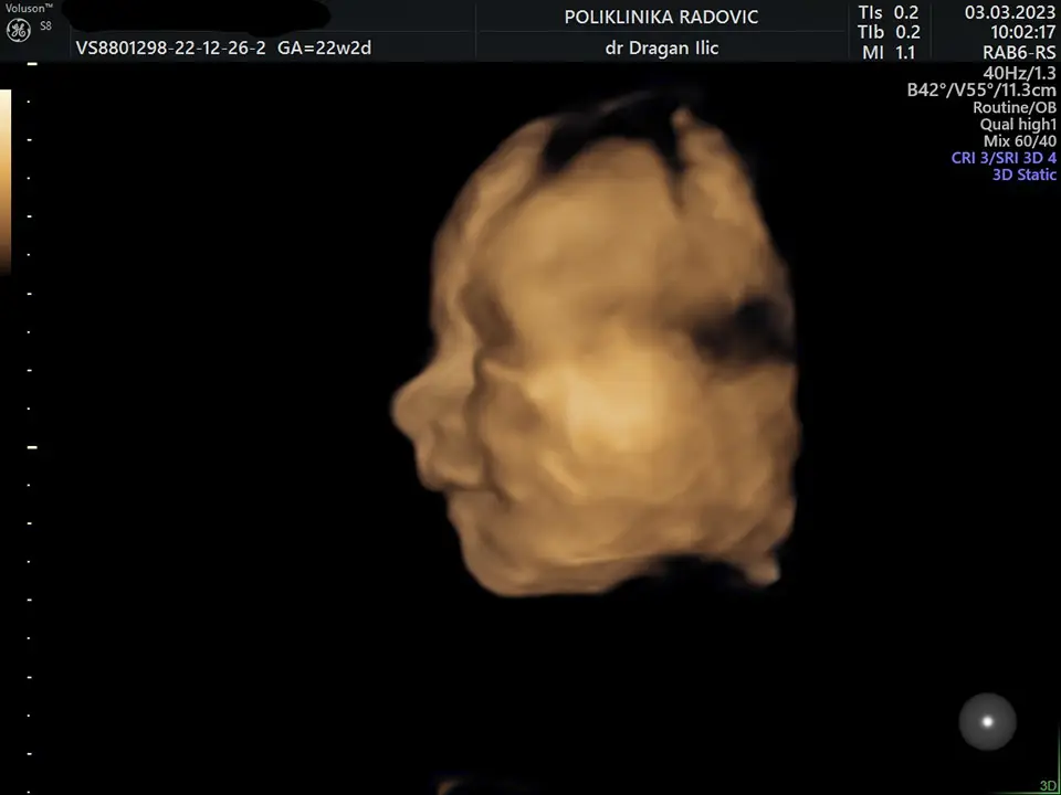
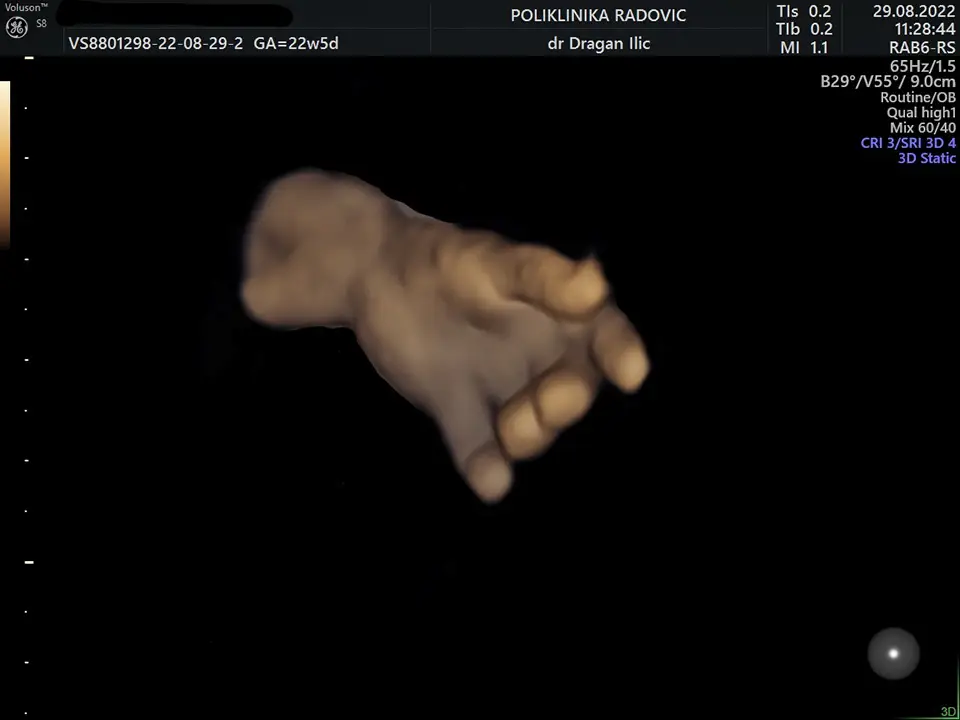
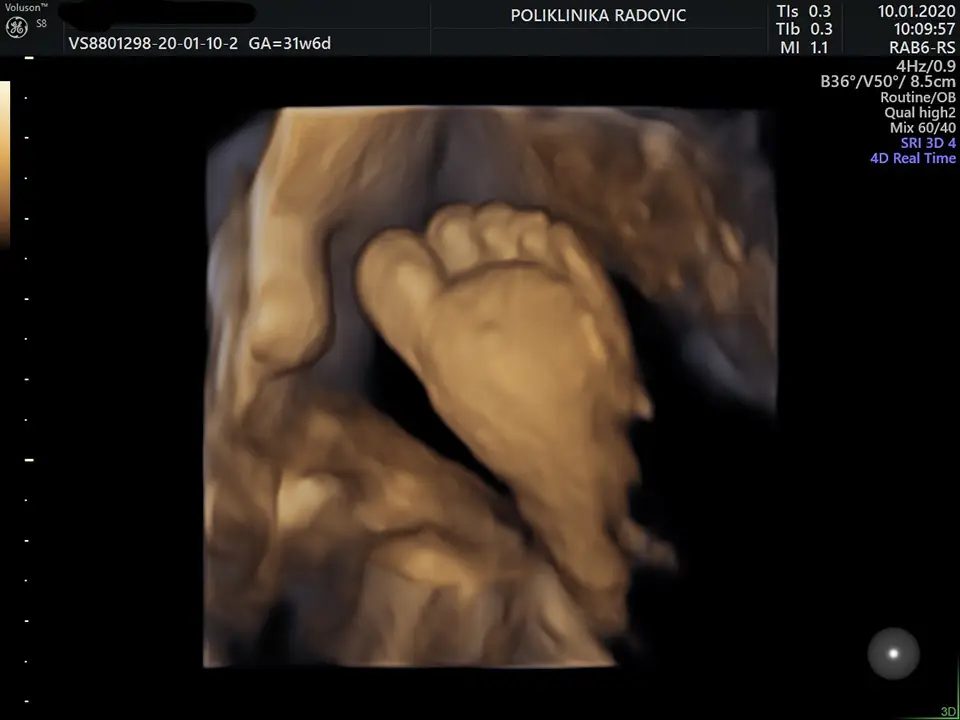
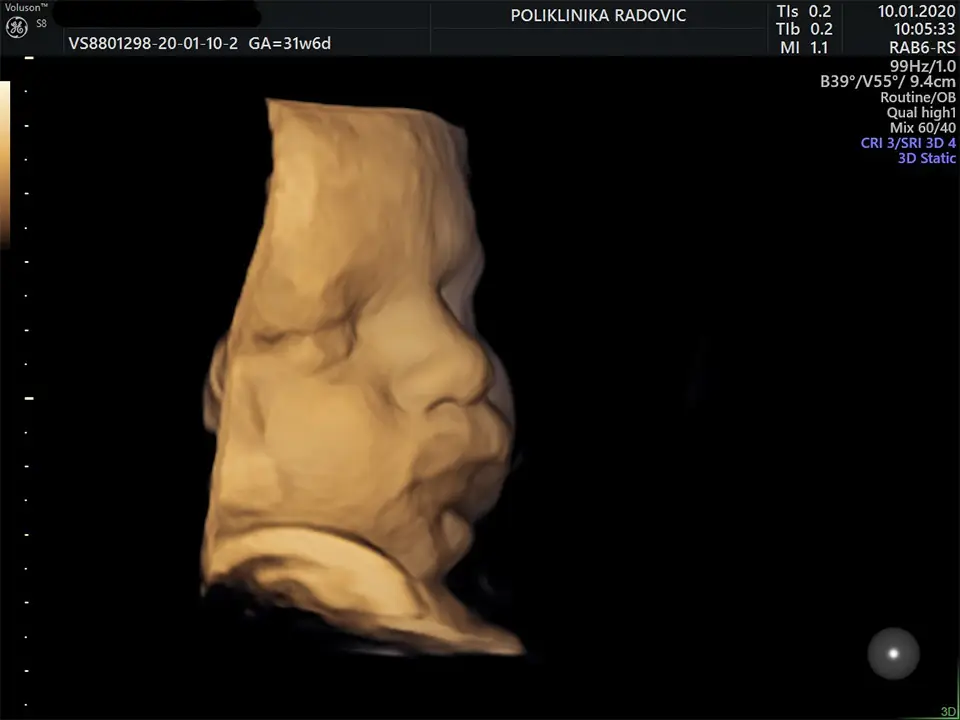
Recently, with the accelerated development of technology, the availability of 3D/4D ultrasound has become a reality. Its importance is that it gives a clear picture of the external morphology of the fetus in three dimensions, in addition to the width and height seen on a standard 2D ultrasound, we also get depth, and if the baby is observed in real time and its movements, then it is a 4D ultrasound.
With this examination, it is possible to obtain extremely clean and clear images of the external morphology of the baby’s head, extremities, abdominal wall, genitals, and spine. With special examination techniques, a 3D view of the internal organs of the fetus is possible.
The role of 3D ultrasound in gynecology and pregnancy.
With a special vaginal 3D ultrasound probe, it is possible in gynecology to observe the uterine cavity, the endometrium, in order to discover the anatomical causes of sterility. A clearer view of endometrial septa, polyps and submucosal myomas is possible.
After all of the above, it should be emphasized that the information obtained by a routine standard 2D ultrasound examination is extremely invaluable. A 3D/4D ultrasound examination cannot replace a standard 2D ultrasound examination, but it can significantly complement it, especially during expert ultrasound examinations. It is best to do it after a second expert ultrasound examination, or if the patient wishes, and if the position of the fetus allows it, it can be done during the entire pregnancy.
Bearing in mind that not every pregnancy is the same, it is usual to perform an average of 5-7 ultrasound examinations during pregnancy, the most important of which are those of 12-14 NG and 20-22 NG. Ultrasound examinations cannot adversely affect the fetus.
Dr. DRAGAN ILIC
A specialist
GYNECOLOGY AND OBSTETRICS


